
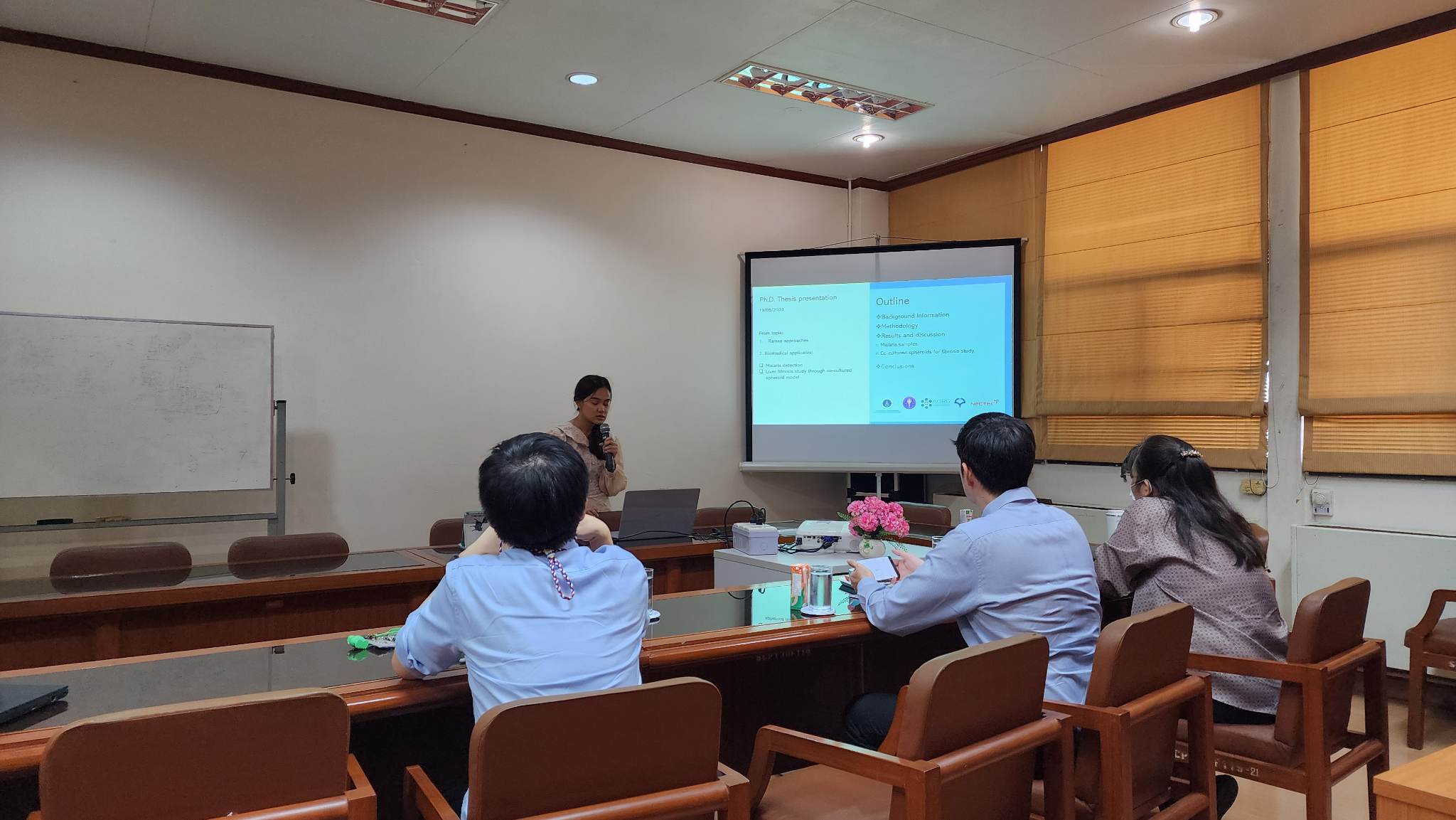
Oral thesis defence examination
ขอแสดงความยินดีกับ นางสาวกันต์กนิษฐ์ คงกลัด นักศึกษาจากกลุ่มวิจัยทัศนศาสตร์ประยุกต์และกลุ่มวิจัยฟิสิกส์ศึกษา ภาควิชาฟิสิกส์ คณะวิทยาศาสตร์ มหาวิทยาลัยมหิดล ผ่านการสอบ “Oral thesis defence examination” เพื่อสำเร็จการศึกษาในหลักสูตร ปรัชญาดุษฎีบัณฑิต สาขาวิชาฟิสิกส์ (หลักสูตรนานาชาติ) โดยได้ทำจิจัยและได้นำเสนอผลงานในชื่อ “OPTICAL TECHNIQUE-BASED LABEL-FREE RAMAN APPROACHES FOR BIOMEDICAL APPLICATIONS: MALARIA DETECTION AND LIVER FIBROSIS IN COCULTURED SPHEROID MODEL”
ABSTRACT
Raman scattering exploits the frequency shift of the wave relative to the incident beam, reflecting the molecular fingerprint of the material. With their non-destructive nature, label-free detection, and simple sample preparation, Raman approaches have been widely used in the study of biomedical samples. This work investigates the applications of Raman techniques in studying malaria and liver fibrosis. Malaria is an endemic disease in tropical countries like Thailand, where the most severe and potentially fatal form is caused by Plasmodium falciparum. In this study, blood samples containing normal red blood cells and Plasmodium falciparum-infected red blood cells were examined on a glass slide and a surface-enhanced Raman substrate (OnSpec chip) using the Raman microscope with the point illumination configuration. Our findings demonstrate a potential of the Raman technique as an alternatively effective method for detecting malaria samples. The intensity of Raman spectra at 747, 1128, 1228, 1372, 1560, and 1620 cm-1, corresponding to hemozoin, the digested product of hemoglobin, was used to distinguish between normal and Plasmodium falciparum-infected red blood cells. Additionally, a separation model based on the PCA-LDA mechanism was developed and analyzed using Raman spectra. The assessments of the blind samples of blood by volunteers revealed that the separation model accurately classified different sample types with up to 80% accuracy.
To expedite the acquisition of Raman spectra in the microscope, the line illumination configuration was utilized, where a line-shaped laser was used to examine the sample area. This allowed for the rapid acquisition of a large number of spectra, resulting in hyperspectral Raman images. This is the first application of the Raman technique to 3D samples for the study of liver fibrosis. Liver fibrosis is an irregular recovery response that can be caused by various complications, including hepatocellular carcinoma (HCC), a leading cause of death in Thailand. HCC can progress to liver fibrosis due to chronic liver damage. In this study, co-cultured HepG2-LX2 spheroids were used as a 3D in vitro liver model, consisting of two distinct cell types. Hyperspectral Raman images were used to distinguish between the different cell types within the co-cultured spheroids. HepG2 and LX2 cells exhibited distinct intensities of cytochrome-related bands and autofluorescence. The culture medium of co-cultured spheroids was supplemented with liver fibrosis activating factors, including fatty acids and TGF-β, two days before observation. TGF-β triggered fibrosis, while fatty acids caused injury to the co-cultured spheroids. Raman images clearly revealed the deposition of collagen and lipid accumulation following the addition of the activating factors. In the two aforementioned applications, the potential of Raman approaches in biomedical applications was demonstrated. Raman spectra allow for the simultaneous presentation of a lot of biomolecule information. There were many spectra gathered, and they can all be preserved for being an updateable database. This Raman approach’s capabilities may allow for future advancement in terms of clinical diagnostics.
ภาพกิจกรรม
ผู้สนใจในงานด้านนี้สามารถติดตามเพิ่มเติมได้ที่นี่
- Kongklad G, Chitaree R, Taechalertpaisarn T, Panvisavas N, Nuntawong N. Discriminant Analysis PCA-LDA Assisted Surface-Enhanced Raman Spectroscopy for Direct Identification of Malaria-Infected Red Blood Cells. Methods and Protocols. 2022; 5(3):49.