Wisdom of the Land

 
Granting Agency

Biophysics Web
   
   
Published Journal
 
Online Resources
 
Dictionaries
 
 
Leptospira

Effects of static magnetic field on growth of leptospire,
Leptospirainterrogans serovar canicola: immunoreactivity
and cell division

Wannapong Triampo a, d, Galayanee Doungchawee b, Darapond Triampo c, Jirasak Wong-Ekkabut a and I-Ming Tang a, d


a Department of Physics, Faculty of Science, Mahidol University, Bangkok, 10400, Thailand
b Department of Pathobiology, Faculty of Science, Mahidol University, Bangkok, 10400, Thailand
c Department of Chemistry, Faculty of Science, Mahidol University, Bangkok, 10400, Thailand
d Capability Building Unit in Nanoscience and Nanotechnology, Faculty of Science, Mahidol University, Bangkok, 10400, Thailand
Received 4 March 2004;  accepted 14 June 2004.  Available online 19 October 2004microscopy;


Abstract

The effects of the exposure of the bacterium, Leptospira interrogans serovar canicola to a constant magnetic field with magnetic
flux density from a permanentferrite magnet=140±5 mT were studied. Changes in Leptospira cells after their exposure to the field
were determined on the basis of changes in their growth behavior and agglutination immunoreactivity with a homologous
antiserum using dark-field microscopy together with visual imaging. The data showed that the exposed Leptospira cells have
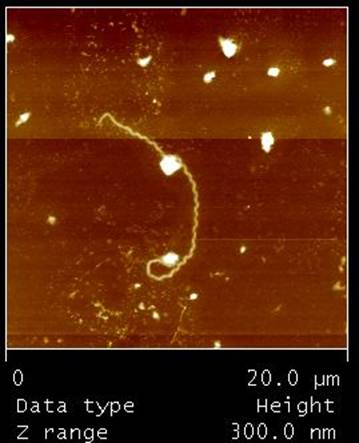
lower densities and lower agglutination immunoreactivity than the unexposed control group. Interestingly, some of the exposed
Leptospira cells showed abnormal morphologies such as large lengths. We discussed some of the possible reasons for these
observations.

Author Keywords: leptospirosis; Leptospira interrogans; magnetic field; dark-field

Selected results (Full paper..(pdf))

----------------------------------------------------------

----------------------------------------------------------


An original atomic force microscopy (AFM)   of
Leptospira interogans serovar canicola taken
using Digital Instrument NanoScope IIIa contact
mode under controlled condition without MF
exposed  with scan size is   and scan
rate is 1 Hz. It  demonstrates a spiral shape
leptospire with approximately .


----------------------------------------------------------


Dark field micrographs of L. interrogans serovar
canicola
exposed to MF for different durations.
The images weretaken at the log phase of each
experimental culture sample
(diluted 1:10 of original). Bars: 100 μm.


----------------------------------------------------------


Dark field micrographs of agglutinated bacterial
cells after reacting with homologous antiserum,
showing complete agglutination (100% reactivity; A)
and 50% agglutination with one-half of free-living
bacterial cells remaining (B).


----------------------------------------------------------


Plots of data shown in Table 1


----------------------------------------------------------


Dark-field micrographs of L. interrogans serovar
canicola taken at the same magnification(×200).
Control sample unexposed to magnetic field;
the leptospires have an approximate length
of 10–20 μm (A) compared with magnetic
field-exposed leptospires (B) with some
cells longer than others. Circles indicate
individual
bacterial cells. Bars: 100 μm.


Table 1. Agglutination characteristics of leptospires after magnetic field exposure for various durations


© 2005 Biophysics Group, Faculty of science, Mahidol University. All Rights Reserved. Contact Webmaster for more information
Last Updated : October 29, 2010