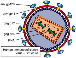
The Human Immunodeficiency Virus (HIV) is a lentivirus which causes the Acquired Immunodeficiency Syndrome (AIDS), a condition in humans in which the immune system begins to fail, leading to life-threatening opportunistic infections . HIV’ size is around 120 nm with its half life approximately 6 hours and its mean reproduction rate around 1010 virions a day[1-2]. In construction, HIV uses the host cell membrane to make its viral envelope. This envelope is covered by gp41 and gp120 surface proteins. Inside the viral envelope is a layer called the matrix, which is made from the protein p17. HIV core is usually bullet-shaped and it is made from the protein p24. Inside the core are three enzymes required for HIV replication call reverse transcriptase, integrase and protease. Also held within the core is HIV’s genetic material, which consists of two identical standards of RNA (see Fig. 1)[3-4].

Figure 1. HIV structure.
Source: http://www.avert.org/hiv-virus.htm
Like all viruses, HIV cannot grow or reproduce on its own. Hence, in order to make new copies of itself, it must infect into the cells of a living organism. After infection to human body, HIV is rapidly going to be spread throughout the circulatory system and lymphatic system to cripple our immunes’ organization. HIV life cycle begins with attachment of the virus to the host membrane which has CD4 protein as the receptors on its surface particularly in CD4+T cells, together with CCR5 (for infecting macrophage) or CXCR4 (for infecting CD4+T cell) as co-receptor to fusion into the host. After entry into the host, the viral RNA genome undergoes reverse transcription, and then the proviral DNA integrates into the host chromosome. After translation, the viral proteins assemble at the cell membrane. At this time, the immature viral particle which contains the RNA genome and viral enzymes egresses the cell. After virion budding, proteolytic processing of the capsid leads to a structural rearrangement of the virion and generates a mature viral particle[5].
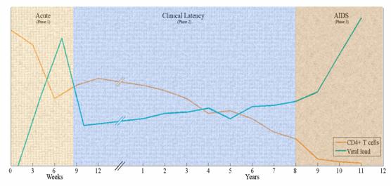
In pathogenesis, HIV distinctly leads to a progressive reduction in the number of CD4+ T cells . Therefore, medical professionals use the CD4 count to refer the stage of infection and to decide when to begin treatment for HIV infected patients. The typically evolution of HIV is divided in three phase pattern shown as follow (see figure 2).

Figure 2. The typical evolution of HIV divided in three phases represents in two time scales (weeks and years). The two lines identify the dynamics of CD4+T cells and viral load, respectively.
- During a few weeks, a transient and dramatic jump of plasma virion level is present with a marked decrease of immune cell count (CD4+T cells), following by sharp decline.
- In the subsequent clinical latency phase, (varying from one to ten or more years, on average eight to ten years) the immune system partially eliminates the HIV virus and the rate of viral production reaches a lower, but relatively steady, sate that varies greatly from patient to patient. Their apparent good health continues because CD4+T cell levels remain high enough to preserve defensive responses to other pathogens. But over time, CD4+T cell concentrations gradually fall.
- An outbreak of the virus (varying from one to two years), together with constitutional symptoms and onslaught by opportunistic diseases, cause death[6].
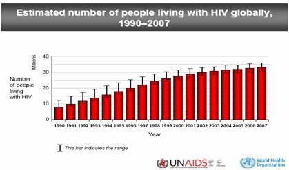
Now a day, HIV infection has shown a high degree of prevalence in population all over the world (see also figure 3) [7]. Because the exact mechanism between HIV dynamics and its interaction against the immune system are not well understood, the HIV infection is still currently incurable. Therefore, it has been widely studied both in the laboratory and with computer modeling in order to understand the different aspects that regulate the virus-host interaction and several treatment strategies such as antiretroviral drugs and vaccine designs[8-12] for many years with the best hope for future control.

Figure 3. Estimated number of people living with HIV globally in 1990-2007.
Source: W.H.O. Unaids, AIDS epidemic update: December 2007. (2007).
Our research group is one that conducted research on this disease for many years by trying to invent and use mathematical models and simulations in which base on, for instance, ordinary and partial differential equation (ODE/PDE) including cellular automata (CA) method. With the hope that we can create the models that are realistic and can be effective in explaining the dynamics of the disease, benefit or inspire a nation knows in the future.
Our ongoing works:
- Developed an ODE based on the relationship between HIV and immune cells such as CD4, CD8 and B cell in tissues and blood to describe the dynamics of HIV.
- Describes the temporal and spatial formation of infected cells propagating in lymphoid tissue by cellular automata model.
References
1. Perelson, A.S., et al., HIV-1 dynamics in vivo: virion clearance rate, infected cell life-span, and viral generation time. Science, 1996. 271(5255): p. 1582.
2. Ho, D.D., Perspectives series: host/pathogen interactions. Dynamics of HIV-1 replication in vivo. Journal of Clinical Investigation, 1997. 99(11): p. 2565-2567.
3. Noble, R. HIV structure and life cycle. AVERTing HIV and AIDS
2009 July 30, 2009; Available from: http://www.avert.org/hiv-virus.htm
4. Mandell, G.L.e.a., Principles and Practice of Infectious Disease. 6th ed. 2004: Churchill Livingstone.
5. Simon, V. and D.D. Ho, HIV-1 dynamics in vivo: implications for therapy. Nature, 2003. 1(3): p. 181-190.
6. Hershberg, U., et al., HIV time hierarchy: winning the war while, loosing all the battles. Physica A: Statistical Mechanics and its Applications, 2001. 289(1-2): p. 178-190.
7. Unaids, W.H.O., AIDS epidemic update: December 2007. 2007.
8. Fauci, A.S., et al., HIV vaccine research: the way forward. Science, 2008. 321(5888): p. 530.
9. Girard, M.P., S.K. Osmanov, and M.P. Kieny, A review of vaccine research and development: the human immunodeficiency virus (HIV). Vaccine, 2006. 24(19): p. 4062-4081.
10. Letvin, N.L., Progress toward an HIV vaccine. Annual Review of Medicine, 2004. 56: p. 213-223.
11. Cohen, J., Shots in the dark: the wayward search for an AIDS vaccine. 2001: WW Norton & Company.
12. Letvin, N.L., Progress in the development of an HIV-1 vaccine. Science, 1998. 280(5371): p. 1875-1880.
Opportunistic infection is an infection by a microorganism that normally does not cause disease but becomes pathogenic when the body's immune system is impaired and unable to fight off infection, as in AIDS and certain other diseases.
CD4+ T cells (also known as T helper cells or effecter T cells) are a sub-group of lymphocytes (a type of white blood cell or leukocyte) that play an important role in establishing and enhancing the capabilities of the immune system. T helper cells are unusual in that they have no cytotoxic or phagocytic activity; they cannot kill infected host cells or pathogens. Their function only active and direct other immune cells to the antigen, and without other immune cells they would usually be considered useless against an infection. Mature T helper cells are believed to always express the CD4 protein on the surface, and are also known as CD4+ T cells. The importance of CD4+ T cells can be seen from HIV in order to treat as having a pre-defined role as helper T cells within the immune system.
----------------------------------------------------------------------------------------------------------